Part of the
4TU.
Federation
TU DelftTU EindhovenUniversity of TwenteWageningen University
4TU.
Federation
NL|EN
Close

4TU.Federation

+31(0)6 48 27 55 61

secretaris@4tu.nl

Website: 4TU.nl

Interview with 4TU's Precision Medicine Team

Thursday, 26 November 2020

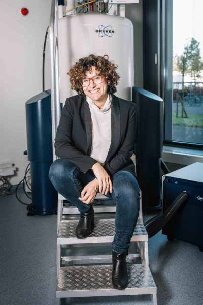
The right treatment, in the right way, at the right time. The 4TU.Federation interviewed Tenure trackers Camilla Terenzi (WUR) and Jelmer Wolterink (UT) on the dream that connects the 4TU Precision Medicine team.

Photos: Dieuwertje Bravenboer

What is the 4TU Precision Medicine programme all about and what’s your role in it?

Jelmer Wolterink (JW): The aim of the Precision Medicine programme is to collect more information from medical imaging techniques, such as MRI, CT and ultrasound. By bringing together all the information extracted from medical images, including the physics that describes how the images were made and analysing it using artificial intelligence (AI), we aim to achieve faster and more accurate diagnoses.

Ultimately, our dream is for patients in the future no longer to be given the one-size-fits-all treatment, but for us to apply exactly the right treatment, in the right way, at the right time. For example, in cases where a patient now has to undergo three scans to detect a tumour, we want to be able to obtain the information we need in one go. This will improve the patient's experience and, eventually, also their quality of life. That in turn will immediately lead to the desired result of saving time for doctors and reducing costs for healthcare.

Short biography Jelmer Wolterink (UT)

Jelmer obtained his PhD in 2017 at Utrecht University (UMC Utrecht) with a thesis entitled Machine learning based analysis of cardiovascular images. After postdocs in the quantitative image analysis group at UMC Utrecht and Amsterdam UMC, he started as an assistant professor in the Applied Analysis group in Twente in March 2020. Jelmer’s research interests are in machine learning for medical image analysis, in particular generative models and geometric deep learning approaches.  

Camilla Terenzi (CT): It’s a dream that we share with the whole team made up of people with different areas of expertise.

Which areas of expertise?
CT: Our consortium includes scientists from various backgrounds, ranging from fundamental physics to computer science and applied mathematics. We bring all of these different pieces of knowledge together. Knowledge about medical imaging techniques, for example. A specific example is an MRI scan of the heart. Can we speed up the scan and also ensure that the image the scan produces still contains the same information as it does now? Or can we be more effective in retrieving information that is currently collected but is left unused? How can we use our expertise to ensure that an image actually shows everything you’re measuring? Could physics be useful here and will it help us to advance diagnostics a step further?

What are you proud of?
JW: A really good example in my view is the MRI scanner that’s set up in the TechMed Centre in Twente. The testing facility in which this MRI scanner is placed is identical to that in hospitals. Scientists from the University of Twente are working with their counterparts at TU Delft, Wageningen University (WUR) and Eindhoven University of Technology (TU/e) to find out how much data you can remove from an MRI scan while still getting a valuable image. By learning where exactly the clinical information is hidden, you could in the future be able to reduce the time that a patient spends in the scanner from the current 45 minutes maybe to just three minutes. As well as benefiting the patient’s well-being, that also greatly improves efficiency! 

“By learning where exactly the clinical information is hidden, you could in the future be able to reduce the time that a patient spends in the scanner from the current 45 minutes maybe to just three minutes.”
Jelmer Wolterink (UT)

There’s also another great project. In it, researchers from the University of Twente, TU/e and TU Delft are using an ultrasound device to measure how microscopically small bubbles – smaller than red blood cells – respond to ultrasound. The device has the sensitivity to detect these bubbles, thereby providing a live image of the  organ perfusion. We intend to use AI to see whether we can further improve these techniques. Applications include ultrafast 3D imaging of blood flow in the abdominal artery or accurately measuring new blood vessel formation in tumour tissue. This information will help vascular surgeons and oncologists to develop better diagnoses and treatment plans that focus on each specific patient. 

Short biography Camilla Terenzi (WUR)

Camilla started as an Assistant Professor in NMR/MRI methods applied to soft matter and food science at the Laboratory of Biophysics of Wageningen University in July 2018. Before that, she did post-doctoral research at Cambridge University (UK), University of Nancy-Lorraine (FR) and KTH Stockholm (SE). She got her PhD in Physics at Sapienza University of Rome on “2D NMR relaxometry in porous media”. Camilla’s research interests are in quantitative NMR/MRI dynamic studies of fundamental and industrially-relevant dynamic phenomena in multi-phase systems.

CT: The development we’re experiencing as scientists can equally well be described as a success. As a result of this joint programme, you discover new avenues of research. For example, although I don’t have a background in medical physics, I’m being encouraged by my fellow researchers to develop that side of myself and engage in discussion with them. Exchanges like that produce lots of new ideas and therefore also achieve a high impact when it comes to peer-reviewed articles for scientific journals. 

And Jelmer also has something to be proud of?
JW: Yes, that's right! Last November, I received a VENI grant from the NWO for EUR 250,000. As a scientist, that gives you the opportunity to develop a new line of research. The money will be spent on improving the risk prediction for rupture of the main abdominal artery, the aorta. The chance of rupture occurs when the aorta widens. Currently, surgical intervention is usually done if this enlargement exceeds 5.5 cm, but rupture can also occur if an aorta is narrower than 5.5 cm. On the other hand, surgical intervention is not without risk and you only want to operate on patients who really need it. My plan is to combine AI and the various imaging techniques in order to achieve a better assessment of the risk of rupture and treat patients in a more targeted way. As well as with doctors, I also work a lot with researchers from other universities, including those in the 4TU programme. This helps to ensure that the knowledge is circulated more rapidly across different specialisations. 

“I’ve worked in Sweden, France and in the UK, but I have never experienced that degree of connection. This country's compact geography enables us to take full advantage of all of the facilities and bright scientists the Netherlands has to offer.”
Camilla Terenzi (WUR)

That kind of knowledge sharing is also characteristic of the 4TU programme?
CT: Yes, certainly! Aside from the collaboration between researchers, the power of this programme also lies in the connections that there are with almost all academic hospitals. That’s another way of increasing the impact of our research. And we need the people from the clinic to make sure we have access to the data required. I’ve worked in Sweden, France and in the UK, but I have never experienced that degree of connection. This country's compact geography enables us to take full advantage of all of the facilities and bright scientists the Netherlands has to offer.

When does this programme end?
CT: The programme started in 2018 and runs until December 2022. But that’s only the end of the beginning: the robust network that we’re currently building will remain. It will prove extremely useful in writing new research proposals in the future. Within our network, we work with talented post-docs, PhD candidates and students. We are the drivers, but they are the driving force! 

The Precision Medicine HTSF programme

The aim of the 4TU Precision Medicine programme is to integrate deep learning, a special kind of artificial intelligence, and medical imaging techniques to raise the level of diagnostics – from a one-size-fits-all approach to a tailored, personalised approach. According to the Precision Medicine team, this is the way to keep healthcare accessible and affordable in the long term. 

The team includes seven Tenure Track candidates, high-performing academics who are given the opportunity to progress to an associate professor position and ultimately to the position of full professor. Some of the scientists have dual roles at medical centres and there is also collaboration with clinical partners as part of the Centres for Medical Imaging in the Innovative Medical Devices Initiative (IMDI): Quantivison (Amsterdam UMC, NKI/AVL), IDII (TU/e, Maastricht UMC+, UMC Utrecht), CMINEN (UT, Radboudumc, UMC Groningen) and Medical Delta (TU Delft, Leiden UMC, Erasmus MC). 


Program leader

Prof. dr. Michel Versluis
University of Twente
Physics of Fluids Group, MESA+ Institute for Nanotechnology, Technical Medical (TechMed) Center
m.versluis@utwente.nl


Tenure trackers

Dr. Camilla Terenzi
Wageningen University & Research
Laboratory of Biophysics, Agriculture and Food Science Group
camilla.terenzi@wur.nl


Dr. David Maresca
Delft University of Technology
Department of Imaging Physics, Faculty of Applied Sciences
d.maresca@tudelft.nl


Dr. Guillaume Lajoinie
University of Twente
Physics of Fluids Group, MESA+ Institute for Nanotechnology, Technical Medical (TechMed) Center
g.p.r.lajoinie@utwente.nl

Dr. Jelmer Wolterink
University of Twente
Department of Applied Mathematics, Technical Medical (TechMed) Center
j.m.wolterink@utwente.nl


Dr. Min Wu
Eindhoven University of Technology
PULS/e, Department of Biomedical Engineering
m.wu@tue.nl


Dr. Sebastian Weingärtner
Delft University of Technology
Department of Imaging Physics, Faculty of Applied Sciences
s.weingartner@tudelft.nl


Dr. Simona Turco
Eindhoven University of Technology
Biomedical Diagnostics lab, Department of Electrical Engineering
s.turco@tue.nl