Medicine is becoming increasingly personalised. One-size-fits-all approaches make way for tailor-made treatments, for instance for cancer and cardiovascular disease. In the 4TU Precision Medicine programme, scientists are working towards this goal by improving medical imaging technologies in a fruitful interaction between science and clinical practice.
Text: Nienke Beintema | Photography: Dieuwertje Bravenboer
No two patients are the same, even if they have the same illness. Whether they have cancer, a cardiovascular or neurological disease, their diseases may be as different as day and night. Tumours in liver and breast tissue, for instance, may share more characteristics than two different breast tumours.
Clinical practice is increasingly tuned towards this reality. Scientists are looking for ways to characterise their patients and their diseases in more and more detail, in order to offer them tailor-made treatments. Not just to target their disease more effectively, but also to limit adverse side effects.
“Previously, if you had cancer, you’d be given a general cytotoxic drug, and hope that this quite drastic approach would mostly harm the cancer”, says cardiologist Thomas Treibel of University College London. “Now, we can use imaging, molecular and hormonal phenotyping to characterise the cancer and then use very specific drugs to target it, in very specific amounts. Our aim is to translate this approach also into cardiology.”
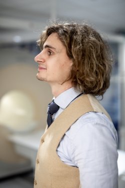
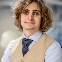
“Imaging is the backbone of this characterisation”, says Sebastian Weingärtner, assistant professor at TU Delft. “I’d argue that imaging puts the ‘precision’ into ‘precision medicine’.” Both scientist have known each other for many years. They’ve been working together in various projects, also during Sebastian’s previous positions.
Imaging as a backbone – is that why 4TU Precision Medicine programme focuses specifically on imaging?
Sebastian: “Yes. If you want to treat a patient effectively, the very first step is an accurate diagnosis. Diagnostic parameters such as blood markers, body temperature and other biometrics are all systemic. They tell you about the state of the entire body. But imaging can tell you exactly what is going on, and where. Imaging is the only approach that can provide that level of detail.”
Thomas: “We cardiologists can make an electrocardiogram, or look for certain peptides that are markers for heart failure. But none of these data show any detail of the actual damage to the heart. Is it on the left or the right side, for instance? Hence, any medication would be quite generic. Imaging such as MRI can give you exact details on scarring in the muscle tissue, or on why a heart valve is malfunctioning.”
Sebastian, you are a physicist. How does physics help to improve MRI?
Sebastian: “The ingenuity of MRI lies in its physics. Much of the differences between the MRI machines that you find at different hospitals lies not in the engineering of the machines themselves, but in the programming of the underlying physics phenomena, and in the data analysis. By working on the underlying physics, we can unlock a wealth of new information. That is what sets MRI apart from other imaging modalities.”
Thomas: “Other techniques, such as ultrasound and X-ray, are about sending and receiving signals as well. But the difference is: with MRI, Sebastian can programme the signal that the magnet sends out, which affects the signal that comes back to the receiver [see text box, ed.]. This is what determines the image that we can then use for diagnostics.”
Sebastian: “An MRI machine can detect a huge amounts of contrasts. We are only starting to scratch the surface of all the information we can extract from these data.”
How does your research reach the clinic?
Sebastian: “When there are new developments in the science behind MRI, hospitals don’t need to replace their machines. They just need to adjust what the machine does, which is relatively easy. What I develop today, could be used in practice next week. The turnaround times are extremely quick.”
Thomas: “When Sebastian writes a new algorithm, we just need to get a software upgrade, and then we’d immediately get an improved picture or more stable image, for instance.”
How does this benefit the patient?
Thomas: “Well, obviously the patient benefits from a faster and better diagnosis, and hence more targeted treatment. But improved MRI also increases patient-friendliness. At University College London, we are looking at using MRI to reduce the need for invasive biopsies in cases of suspected prostate cancer. There is a cost aspect, but definitely also improved patient comfort and safety. There are huge patient benefits.
Another example: in case of MRI of the heart, the patient has to lie flat and hold his breath during scanning. With increased picture stability, scanning is considerably faster. Sebastian wrote a sequence, for instance, that allows us to quantify tissue changes in the heart in a single breath hold.”
How important is cooperation between science and clinic, like in the 4TU programme?
Sebastian: “Realistically, advancing MRI technology is not on the mind of your average cardiologist. Our tasks as scientists is to make a contribution and then go out and distribute it among hospitals, if the technique proves to be beneficial.”
Thomas: “But to prove that it is beneficial, you need to gather evidence. Not just of economic benefits, but also of benefit to patients, both in the short and the long term. To gather this evidence, you need an effective interface between academic institutions and the clinic. Not just to have an optimal exchange of knowledge, but also just to have sufficient patient numbers. For this, you need an international network. I’ve been working with Sebastian for many years now, and also with other scientists in Europe and elsewhere in the world.”
Sebastian: “At 4TU, we cooperate closely with clinical centers, such as Holland Proton Therapy Centre, where I perform most of my MRI research, as well as with academic hospitals in the Netherlands, and many clinical partners abroad. The 4TU programme fits into this larger international network and benefits from it. But it works both ways: the programme also helps to advance the national research agenda. It groups together scientists from disciplines that wouldn’t usually cooperate.”
And what about the interface with industry?
Sebastian: “We also work closely together with large industry players, like Philips. Here, too, the benefits work both ways.”
Thomas: “And hospitals benefit too. If we can make our scans faster and better, and generate more data, we can improve patient health and comfort and generate a better diagnostic yield in the same amount of time. This will become increasingly important, as we expect the disease burden to keep rising due to the ageing population and post-covid disease, for instance. We’ll need to keep speeding up and improving our diagnostics. A faster, physics-driven approach can do that.”
Which are the main challenges on the road to clinical application?
Sebastian: “As physicists, after having shown on a small scale that our innovations can make a difference, we need to show that our results have a broader applicability, and are reproducible across many different centres. That kind of research is a lot harder to finance. That is where programmes like 4TU Precision Medicine can make a difference.
And there are also considerable regulatory burdens. So there are always translational gaps that we still need to bridge. It’s a long and strenuous road.”
Are you optimistic in this regard?
Thomas: “It is not a question of optimism or pessimism. It is a question of time. The technique needs to be so solid that we can make life-and-death decisions based on it. So we need to be patient. Getting a new technique implemented in all the hospitals in the Netherlands, for instance, is a daunting task. But eventually it can make a difference for hundreds of thousands of people, so it is definitely worthwhile. And in the meantime, we are already using increasingly advanced imaging techniques in our patients, delivering more and better quality information and choosing more personalised treatments.”
Sebastian: “Oh, definitely. Every small innovation is already making a difference for patients. Advanced MRI is a clinical workhorse. It is not something that we tinker up in our lab and maybe three people are using it. Its application is speeding up in clinical settings around the world. Where we stand at the moment is quite staggering.”